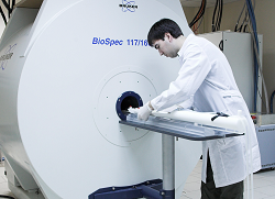
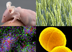
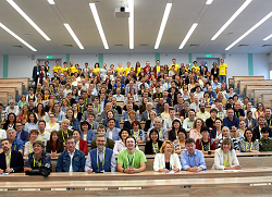
![]() ![]() ![]() |
Laboratory of Biochemical Genetics of AnimalsHead S.M.Zakian, Dr.Biol.Sci., Prof. |
||
Structure of the Xist gene in common voles.
Progression of Xist RNA localization through the cell cycle in the vole M. transcaspicus. DNA/RNA FISH analysis of vole cells showing Xist RNA detected with FITC (green) and heterochromatin block repeat DNA detected with TR (red) illustrated on cells at (a) interphase, (b) prophase, (c) metaphase, (d) anaphase, (e) telophase and (f) daughter cells in the early G1.. DNA counterstained with DAPI
Localization of Xist RNA on the rodent metaphase
X chromosomes.
Assignment of the Xist, Pgk, G6pd, Gla and Hprt genes in M. arvalis (i), M. kirgisorum (ii), M. transcaspicus (iii), M. rossiaemeridionalis (iv) and M. agrestis (v) to specific regions on the X chromosomes by FISH. Top: representative X chromosomes with dual signals from a pair of gene probes. Bottom: computer-generated, G-like banding of the same chromosomes based on DAPI staining. |
The primary objective of the laboratory is to conduct research on the molecular mechanism of X chromosome inactivation in female mammals using the model of interspecific hybrids between common voles of the Microtus genus. The model is unique because the heterochromatin blocks are specifically located on the X chromosomes and also because their inactivation deviates from random in certain cross combinations. The model has advantages over other models allowing to elucidate the possible X-inactivation mechanism. To clarify the causes of the observed phenomenon, a combination of research is applied. It includes localization of the molecular structure of the Xist (X Inactive Specific Transcript) gene in the studied voles and analysis of its expression during development. Research is being carried out on the regional mapping of a number of X-linked genes, the organization and evolution of the common vole genome with the ultimate goal of providing a basis for molecular studies. Search for the X-specific repetitive sequences and proteins involved in the compaction of the X chromosome would also contribute to a better understanding of the mechanisms of the observed phenomenon. The Xist gene sequences and their adjacent 5' regions were completely sequenced in all the four common vole species. Thus, important information about the functionally significant elements in Xist RNA and the Xist promoter was obtained. A close association between Xist RNA and the X chromosome material was established throughout the interphase-metaphase. It was shown for the first time that Xist RNA does not bind homogeneously to the X chromosome material, being located in specific light G-segments, and also that there is no association of Xist RNA with the X-linked heterochromatin. Chromosomal localization of 5 unique X-linked Xist, Gala, G6pd, Hprt, and Pgk was performed using fluorescent hybridization in situ. Thorough studies on the structure of the Xist gene, as well as mapping of this and the other X-linked genes, provided a breakthrough in clarification of the molecular structure of Xist in voles. The results now underlie further studies of the mechanisms of X-inactivation in hybrid voles. It is suggested that the genetic differences producing non-random inactivation of the parental Xs in voles most probably result from elements that are responsible for the chromosome to be inactivated. This chromosome may be chosen at the level of stabilization of one of the Xist transcripts. It is expected that search for differences in the structure of the promoter regions and alternative spliced Xist transcripts in vole species would allow to identify the functionally important regions involved in switching over the transcript from the unstable to the stable state. These studies would advance us in understanding the role of the Xist gene in the regulation of X-inactivation events in mammals. Research efforts are being applied to obtain different cell systems for studying nonrandom X-inactivation in vitro. It is intended to produce embryonic stem cell lines in voles and their hybrids in certain cross combinations, as well as hybrid cells by fusion of mouse ES cells with vole somatic cells and microcells. |
||
|
| |||